Cardiac output is a measurement of how much blood is being pumped by the heart, it is calculated by multiplying the speed at which it is pumping by how much it pumps each stroke.
Cardiac output= heart rate x stroke volume.
Tuesday, 28 January 2014
Wednesday, 15 January 2014
Myogenic stimulation of the heart and transmission of a subsequent wave of electrical activity. Roles of the sinoatrial node (SAN), atrioventricular node (AVN) and bundle of His.
Myogenic stimulation means that the heart is caused to move because of signals that originate inside of it. In normal muscles movement is caused by signals from the CNS.
The signals come from the sinoartrial node (SNA) which is sometimes refered to as the pace maker. Although the signals for the cardiac cycle are generated in the heart, the pace is controlled by signals from the brain.
The signals come from the sinoartrial node (SNA) which is sometimes refered to as the pace maker. Although the signals for the cardiac cycle are generated in the heart, the pace is controlled by signals from the brain.
- SNA sends electrical activity accross the top of the atria (they contract)
- The atrioventricular septum is non-conductive tissue which stops the signal going down the sides.
- The signal that travelled down between the atria reaches the atrioventricular node (AVN).
- The AVN delays the signal (so that the atria fully contract) before transmitting it again.
- The electrical activity now travels down the bundle of His.
- The signal reaches the bottom of the ventricles where it causes them to contract.
Its important that the ventricles contract upwards so that the blood is being pushed towards the semi-lunar valves.
It is also of importance that the AVN delays the signal long enough for the atria to finish their contraction- so that the most blood possible goes into the ventricle before it starts contracting and closes the atrio-ventricluar valves.
Pressure and volume changes and associated valve movements during the cardiac cycle. Candidates should be able to analyse and interpret data relating to pressure and volume changes during the cardiac cycle.
Diastole
The ventricle relaxes, decreasing its pressure. This causes the blood which it just pushed into the aorta/pulmonary (left/right) flow back towards the heart trying to get to the low pressure area: the blood pushes the semi-lunar valve shut (making the second noise of a heart beat 'dub')
Blood flows from the atrium into the ventrical. This happens because there is a lower pressure in it: 1. because it has no blood in and 2. because it has just relaxed.
The blood going through pushes the atrio-ventricular valves open.
The blood going through pushes the atrio-ventricular valves open.
Atrial systole
The atrium contracts: so there is more pressure on the blood. It does this to push the remaining blood into the ventricle that didn't flow in during diastole.
Ventricular systole
Ventricles contract- more pressure- pushing blood through the semi-lunar valves, out of the heart.
When they contract the blood pushes against the atrio-ventricular valve, which pushes it shut (this makes the first sound of a heart beat 'lub')
When they contract the blood pushes against the atrio-ventricular valve, which pushes it shut (this makes the first sound of a heart beat 'lub')
http://library.med.utah.edu/kw/pharm/hyper_heart1.html
.jpg)
Bump- atrium contracts.
Drop- blood is pushed into ventricle.
Gradual rise- blood fills the atrium.
Drop- blood flows into ventricle.
Gradual rise- blood continues to flow in and through into the ventricle.
Ventricle (left) pressure:
First small bump- blood is pushed in by atrium.
Massive bump- the ventricle contracts.
Fall with aorta line- blood has been pushed out.
Fall after the aorta line- ventricle relaxes.
Gradual rise- pressure is below that in the atrium so blood rushes in.
When the ventricle pressure rises above that in the atrium, the atrio-ventricular valve is pushed shut.
When the ventricle pressure falls below that of the atrium, the atrio-ventricular valve opens.
Ventricle (left) volume:
Initial rise- blood is pushed in from the atrium.
Fall- blood is pushed out into the aorta.
Plateau- both valves are closed so no blood is moving in or out (volume can't change.)
Increase- blood flows in from the atrium.
Aorta:
When the pressure of the ventricle meets that of the aorta the semi-lunar valve opens.
Big bump- blood is pushed in from the ventricle.
The ventricle pressure drops below that of the atrium, back flow shuts the semi-lunar valve.
Small bump- the elasticity of the walls brings them in before...
Gradual fall- the walls of the aorta relax.
Valves are pushed open because the pressure has been made grater in one chamber than it is on the other side of the valve, so blood tries to go through it into a lower pressure area, pushing it open.
The gross structure of the human heart and its associated blood vessels in relation to function.
The heart has four main areas: two atriums and two ventricles.
Veins always going into the heart and arteries away from it.
Atrium means entrance hall in latin.
Pulmonary means to do with the lungs.
- Blood first enters the heart into the right atrium from the vena cava;
- passes through the right atrio-ventricular valve into the right ventricle;
- leaves through a semi-lunar valve into the pulmonary artery;
- goes past the lungs in capillaries where it is oxygenated;
- then re-enters the heart through the pulmonary vein into the left atrium;
- passes through the left atrio-ventricular valve into the left ventricle;
- then out through the left semi-lunar valve into the aorta (which takes it to the body.)
 |
| wikibooks |
Veins always going into the heart and arteries away from it.
Atrium means entrance hall in latin.
Pulmonary means to do with the lungs.
The mechanism of breathing.
When volume is increased pressure is decreased.
Air rushes into areas of lower pressure.
So increasing volume in the lungs brings air in:
Breathing in (inspiration)
Diaphragm contracts: flattens- increasing volume.
Intercostal muscles contract: pulling up and out- increasing volume.
When volume is decreased it increases pressure.
Air rushes to areas of lower pressure.
So if volume is decreased in the lungs air will rush out:
Breathing out (Expiration)
Diaphragm relaxes: becoming a dome shape and pushing up- decreasing volume.
Intercostal muscles relax: pushing the ribcage down and in- decreasing volume.
.jpg)
Pulmonary ventilation as the product of tidal volume and ventilation rate.
Tidal volume is the amount of air breathed in. Measured in decemeters cubed.
Ventilation rate is the number of breaths taken in one minute. Measured in minutes to the power of -1.
If you times these two things together you get a measure of pulmonary ventilation in dm3min-1.
Ventilation rate is the number of breaths taken in one minute. Measured in minutes to the power of -1.
If you times these two things together you get a measure of pulmonary ventilation in dm3min-1.
The exchange of gases in the lungs.
Oxygen diffuses from the alveoli- where it is in high concentration- into the capillaries- where it is in low concentration.
Carbon dioxide diffuses from the capillaries- high conc.- into the alveoli- low conc..


Saturday, 11 January 2014
The essential features of the alveolar epithelium as a surface over which gas exchange takes place.
Epithelila cells make up the walls of alveoli.
The alveoli are surrounded by capillaries, this means there is a constant flow of blood that takes oxygen away from the area of diffusion, and the lungs bring a constant supply of oxygen to be diffused on the other side; the opposite is true for CO2- this keeps a big difference in concentration gradient to speed up diffusion.
Both alveoli and capillaries have very thin walls making the diffusion distance short (so diffusion happens more quickly.)
Because there are so many alveoli there is a very large total surface area fro gasses to diffuse through (quicker diffusion.)
There is mucus lining the alveoli which helps gasses to diffuse across.
The gross structure of the human gas exchange system limited to the alveoli, bronchioles, bronchi, trachea and lungs.
Once air is breathed in through the mouth or nose it travels down the trachea. The trachea splits into two- one going into the left lung and one going into the right lung- these pipes are called bronchi. Each bronchus will then divide further into many bronchioles: each ending in a sac called an alveoli.
The trachea and bronchi have walls of muscle that are supported by cartilage. The cartilage is in partial rings so that the tubes can be moved in any direction. Cilia on the walls move mucus out of the breathing system and into the stomach.
The trachea and bronchi have walls of muscle that are supported by cartilage. The cartilage is in partial rings so that the tubes can be moved in any direction. Cilia on the walls move mucus out of the breathing system and into the stomach.
 |
| wikibooks |
Thursday, 9 January 2014
The use of oral rehydration solutions (ORS) in the treatment of diarrhoeal diseases. The applications and implications of science in developing improved oral rehydration solutions; ethical issues associated with trialling improved oral rehydration solutions on humans.
Diarrhoea leaves people dehydrated and with depleted amounts of necessary salts, as these things are lost in faeces. Cholera is an example of an illness that causes diarrhoea.
Oral rehydration solutions (ORS) are used to rehydrate and replenish key molecules.
ORS is a mixture of water, salts and sugars. A simplified ration is 1 litre of water to six teaspoons of sugar to half a teaspoon of salt. The water is often boiled to sterilise it.
- Sodium is needed because it goes into the cells and brings the water potential back down, this means water will move by osmosis back into the cells, and from there into the blood.
- Sodium has to enter the cells through co-transportation (because the normal carrier proteins are pumping sodium out) with glucose- this means that glucose is also needed.
- Other salts that make ORS isotonic; so that when people are drinking it their cells do not absorb all of the water and burst.
To work out the right proportions of salts and liquids, a lot of testing had to be done. This involved trialling early mixtures on humans to note the side effects and make appropriate changes. In the process many peoples conditions were worsened due to having too much of a certain molecule: too much glucose in one trial resulted in more water being drawn into the intestine and the diarrhoea was worsened.
Some claim that it is immoral to jeopardise peoples health by giving them un-trialled solutions; however, by trying out the solutions on people, scientists were able to refine ORS to create an effective treatment that saves many lives.
The specification wants you to say it contains glucose and:
Sodium (ions) / potassium (ions) / chloride (ions) / citrate (ions);
The specification wants you to say it contains glucose and:
Sodium (ions) / potassium (ions) / chloride (ions) / citrate (ions);
Cholera bacteria produce toxins which increase secretion of chloride ions into the lumen of the intestine. This results in severe diarrhoea.
Toxins produced by the cholera bacteria effect the epithelial cells of the intestine- this is because it is complimentary to the receptors that only these cells have.
One half of the toxin binds to a receptor on the surface of the cell- this gives the other half access to the cell, so it can affect the producer of cAMP (that being a signal used within the cell). The signal causes a protein channel to transport chloride ions out of the cell.
The effected cells are in the intestine, which chloride ions will now begin to flood into. The chloride ions lower the water potential in the intestine so water moves into it (along the gradient from high to low.) The intestines are where faeces is made, so when an effected person defecates it has a very high water content- diarrhoea. Because there is a lot of water in the intestine, molecules like salts will move into the intestine too (where they are in lower concentration) meaning that they too are lost in faeces.
The cholera bacterium as an example of a prokaryotic organism. The structure of prokaryotic cells to include cell wall cell-surface membrane, capsule, circular DNA, flagella and plasmid.
The bacterium that causes cholera is a prokaryotic organism as are all bacteria.
These cells contain fewer organelles than an animal cell, and have some other features.
They have a cell-surface membrane, but they also have a cell wall like a plant: they have another layer outside of this to offer additional protection called a capsual.
The DNA in these cells is not contained within a nucleus- it is just a scrunched up ring called circular DNA.
A flagella helps bacteria move- often associated with sperm cells, but they are also important for cholera bacteria to get through the mucus lining to the epithelial cells in the small intestine.
Plasmids are small sections of DNA that are separate from the main circular DNA and replicate separately from it. They can carry genes that the bacteria didn’t always have and can be transferred between bacterium.
Absorption of the products of carbohydrate digestion. The roles of diffusion, active transport and co-transport involving sodium ions.
Carbphydrates are digested from polysaccharides into monosaccharides. These monosaccharides are absorbed into the blood stream. This happens in the small intestine.
Monosaccharides are moving from the lumen (interior) of the small intestine into the epithelial cells of the small intestine (lining) into the blood in the capillaries. There is a good blood flow which takes away the monosaccharides in the blood. So the highest concentration is in the intestine and lowest in the blood making a concentration gradient so monosaccharides can diffuse into the blood stream.
Not all of the monosaccharides in the intestine can be absorbed by diffusion because it relies on the concentration gradient being correct.. so active transport might be used. Monosaccharide molecules can travel into the epithelial cell by a co-transport protein carrier, but only if they are with sodium ions. To make sure sodium ions are travelling into the epithelial cells, sodium ions are transported out of them on the opposite side (into the blood) so that there is a lower concentration in the cells than the lumen: this means that sodium ions will be travelling into the cells (along the concentration gradient). When they travel in, they bind to the co-transport protein, a monosaccharide now also binds to the protein changing its shape so it can release the molecules on the other side. Now the monosaccharide is in the cell facilitated diffusion is used to transport it out the other side into the blood stream.
Wednesday, 8 January 2014
The role of carrier proteins and the transfer of energy in the transport of substances against a concentration gradient.
Intrinsic proteins in the plasma membrane act as carriers, transporting substances across the membrane.
A substance will bind to the carrier protein. The protein then hydrolyses an ATP (energy) molecule: binding to the phosphate product and using the energy from breaking the bond to change shape. This new shape is one that allows the substance to be released on the other side of the membrane.
Often, now that the carrier is open on the other side of the membrane, it will bind to molecules from there so that when it returns to its original shape, they are released over there. After the carrier protein gone back to it original shape, the phosphate ion is let go.
When the ATP is broken up ADP is produced as well as a phosphate ion. They can both be reused, and made back into ATP in respiration.
Seriously watch this, it shows the process perfectly:
http://highered.mcgraw-hill.com/sites/0072495855/student_view0/chapter2/animation__how_the_sodium_potassium_pump_works.html
Osmosis is a special case of diffusion in which water moves from a solution of higher water potential to a solution of lower water potential through a partially permeable membrane. Recall of isotonic will be expected.
Osmosis is a similar movement to diffusion, however given concentration is how much of something is dissolved in water, it is not talked about in terms of concentration. Instead we talk about water potential (Ψ)- referring to how much is dissolved in the water.
0Ψ is pure water, it has nothing dissolved in it. The lower the water potential, the more things dissolved in it. -1Ψ is water with some impurities, -2Ψ has more e.c.t.
Water will move from areas with a high water potential, to areas with a lower water potential, through membranes that are semi-permeable (allow some things through.)
An isotonic solution is one with the same things and amounts dissolved in it as there are inside cells. Cell tissue is isotonic; this means that it will have the same water potential as the cell, and water will not move out of the cell- dehydrating it- or into the cell- bursting it.
Use the fluid-mosaic model to explain appropriate properties of plasma membranes.
Receptors enable the cell to detect hormonal messages;
Enzymes aid chemical reactions outside the cell;
Protein channels facilitate diffusion across the membrane;
Carrier proteins are involved in facilitated diffusion and active transport;
The phospholipid bilayer is semi-permeable so lets water and other substances diffuse in and out.
The role of carrier proteins and protein channels in facilitated diffusion.
Facilitated diffusion is similar to normal diffusion in that molecules are moving from an area of high concentration to an area of low concentration and they are both passive, this means they require no energy. Facilitated diffusion is faster and for molecules that are too big to fit through the plasma membrane, or can't get through because of their charge. Glucose and amino acids both diffuse through the cell membrane in this way.
Intrinsic proteins in the fluid mosaic transport molecules from one side to the other:
Intrinsic proteins in the fluid mosaic transport molecules from one side to the other:
Carrier proteins chemically bond to a specific molecule, when they do this they change shape, the new shape enables the molecule to be released on the other side of the membrane;
Protein channels are passages through the membrane which molecules can pass through if they are the right size to fit through or if they have the right charge to be attracted. Some have gate like mechanisms, by which the protein will allow molecules through as a reaction to stimuli specific of molecule it wants to carry.
Friday, 3 January 2014
Diffusion is the passive movement of substances down a concentration gradient. Surface area, difference in concentration and the thickness of the exchange surface affect the rate of diffusion.
Substances will move from an area of high concentration to an area of low concentration (diffusion.) This often happens when two different environments are connected by a membrane e.g. inside a cell and outside a cell.
A bigger surface area increases the rate of diffusion because there are more opportunities for substances to move across a membrane.
The bigger the difference in concentration the faster the diffusion.
Thickness of the exchange surface affects how far substances have to travel to diffuse into a space: the thicker the surface the longer it takes.
The role of the microvilli in increasing the surface area of cell-surface membranes.
Microvilli protrusions of the cell surface membrane which are tightly packed- spread out they would cover a larger surface area but because they are in a wavy line they don't take up much space but do increase the surface area a lot.
 |
| jornalofillustratedscience |
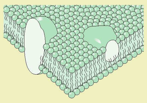
The arrangement of phospholipids, proteins and carbohydrates in the fluid-mosaic model of membrane structure.
The fluid mosaic model is the name for the cell-surface membrane and all the stuff in it; it is knows as fluid because the things in it can move around.
Phospholipids
The phosphate group is attracted to water (hydrophilic), where as the other end of fatty acids are not (hydrophobic)- this means that phospholipids will form a layer on water:
.jpg)
Phospholipids come together so that their tails are facing in and their heads facing out in contact with water. This is called a phospholipid bilayer and is what the cell-surface membrane is made of.
Proteins
Phospholipids
The phosphate group is attracted to water (hydrophilic), where as the other end of fatty acids are not (hydrophobic)- this means that phospholipids will form a layer on water:
.jpg)
 |
| studyblue |
Intrinsic proteins go from one side of the bilayer through to the other. Some are enzymes and others are protein channels which carry substances across the membrane.
Extrinsic proteins are embedded in but do not span the membrane. Some act as structural support; others join with glycolipids to be receptors.
Intrinsic right, extrinsic left:
 |
| kscience |
Carbohydrates
Carbohydrates bond with lipids and proteins to form receptors. The carbohydrate branch acts as a recognition site in glycoproteins, as does the whole glycolipid, to detect chemicals like hormones and toxins.
 |
| malebolge |
The emulsion test for lipids.
Take 2ml of the sample and 5ml of ethanol, then shake- this should dissolve any resent lipids.
Then add 5ml of water and shake again to mix.
If the mixture turns cloudy it is because water is being refracted as it changes between the density of water and lipids: so the cloudy appearance indicates the presence of lipids.
Then add 5ml of water and shake again to mix.
If the mixture turns cloudy it is because water is being refracted as it changes between the density of water and lipids: so the cloudy appearance indicates the presence of lipids.
In phospholipids, one of the fatty acids of a triglyceride is substituted by a phosphate group.
A phospholipid is made up of fatty acids, glycerol and a phosphate group: PO4(3-).
.jpg) |
| Phosphate group; glycerol; fatty acids |
The R-group of a fatty acid may be saturated or unsaturated.
Fatty acids are made up of oxygen, carbohydrate and hydrogen.
In some all of the bonds are being used to join together the separate elements- these are called saturated.
In others not all of the possible bonds that can be made have been, these are unsaturated; this happens because carbons are double bonded (these can be made into single bonds freeing up another space.)
If a fat is unsaturated, its double bonds will cause kinks in the chain- this means that the fats cannot line up and compact into a solid, so they are liquid at room temperature.
Glycerol and fatty acids combine by condensation to produce triglycerides.
A triglyceride is a neutral fat, is formed when one glycerol bonds to three fatty acids.
An 'ester' bond is formed by a three stage condensation reaction.
An 'ester' bond is formed by a three stage condensation reaction.
.jpg) |
| Glycerol; fatty acids; triglyceride; water molecules |
Principles of cell fractionation and ultracentrifugation as used to separate cell components.
Cell fractionation is splitting cells up into its organelles.
- The tissue is chopped up and up into a ice cold, isotonic, buffer solution.
- This is then put in a blender to break open the cells which is called 'homogenisation'.
- The 'homogenate' is then filtered to get rid of debris like connective tissue.
- The mixture is spun on a centrifuge, the densest organelle will collect at the bottom.
- The separated bit at the bottom, the 'pellet' is left in the tube when the homogenate on top which is called the supernatant is poured off into a new tube.
- This new tube is span again to collect the next densest organelle- this is repeated to collect the desired organelles, with the speed increasing each time.
In step one the liquid is cold to slow down enzymes (that might have been freed from lysosomes) so that they don't digest the organelles. It is isotonic to maintain a normal water potential thereby preventing organelles from bursting with water! Buffer solution maintains the PH so that it is appropriate for the organelles.
There are rules on how fast and long you have to spin the centrifuge to get the desired organelle relating to the order of density. From most dense to least the order of these key organelles goes: nucleus; mitochondria; lysosomes; ribosomes.
The principles and limitations of transmission and scanning electron microscopes.
Transmission electron microscopes (TEM) shine a beam of electrons through a slice of stained specimen, some electrons are absorbed and others travel through; the pattern makes a photomicrograph on a fluorescent screen which records a magnified image of the specimen. The electron beam is manipulated by electromagnets:
.jpg)

.jpg)
Scanning electron microscopes (SEM) shin a beam of electrons on the specimen, but instead of going through they scatter off the surface- the electrons are collected and the pattern amplified to give a 3D image of the specimens surface. SEMs have a very complex system but this diagram shows some of the basic components:

TEM SEM
Maximum resolution: 1nm 10nm
Maximum magnification: 250000x 100000x
Image: 2D 3D
In both case the specimen must be dead because it is done in a vacuum. If there was air present the electrons would reflect off of it not the specimen.
TEM requires a complex staining process, and for the specimen to be cut up into extremely small pieces so the electrons can get through. Often TEM show up random artefacts (which are areas on the image that don't really exist) which are a result of the way the specimen was prepared.
The difference between magnification and resolution.
Electron microscopes can magnify objects many times to a good resolution.
Magnification is how many times the image has been enlarged.
Resolution is how clear the picture is. If resolution is 1nm it means that if two objects are one nanometer or more apart the microscope will show them as two different objects; if they are any closer than that they will look like one object.
Waves are only affected by objects that are bigger than half of their length: so big wavelength will only get diffracted by a relatively big object in the way- think of the sea with massive waves- if you put a twig in front of it the water would not be disturbed, you would have to put a tree in front of it to make any difference.
A small wavelength will be diffracted by tiny objects in the way: so the ripples from a raindrop would be affected by a twig. Electrons have a very small wavelength and so are effected by tiny objects- this means that more detail shows up in an electron microscope where the waves have been disturbed than it does on a light microscope (with a bigger wavelength.)
Candidates should be able to apply their knowledge of organelles in explaining adaptations of other eukaryotic cells.
A cell will have organelles to suit its job. The ultrastructure of a cell is just the detail inside it- so its types and amounts of organelles.
Here are some examples of where having more of an organelle fits a cells function:
Here are some examples of where having more of an organelle fits a cells function:
- Mitochondria produce energy (ATP) so are beneficial in muscle cells which need energy for movement and epithelial cells which need energy for active transport.
- Endoplasmic reticulum produces protein (among other things) and so is needed in large amounts for cells that secrete enzymes like the epithelial cells and liver cells
- The same is true for ribosomes which will increase with ER as it covers the surface
- Golgi apparatus enables a cell to egest products like enzymes so would again be helpful in secretory cells; it also produces lysosomes which are needed a lot in phagocytic cells like white blood cells
- Microvilli are helpful in cells that need to absorb things as they increase the surface area, hence why there are so many in the epithelial cells
There can also be adaptations in the organelles- for example in a cell that needs a lot of energy the mitochondria may have more cistae so there is more respiration can happen.
The appearance, ultrastructure and function of the nucleus.
Nucleus:
- Nuclear envelope
- Double membrane
- Controls the movement of substances in and out of the nucleus
- Can have ribosomes on the surface
- Can be continuous with (joined/next to) endoplasmic reticulum
- Contains chromatin
- DNA for the cell
- Form chromosomes take when the cell is not dividing
- Nucleolus
- Makes rRNA (coding for ribosomes)
- Assembles ribosomes
- Makes mRNA (coding for proteins) and controls protein synthesis
- Nucleoplasam
- Viscous liquid
- Molecules and organelles of the nucleus suspended in it
- Holds the structure of the nucleus
 |
| thelogos.co.uk |
.jpg) |
| Not to scale |
The appearance, ultrastructure and function of microvilli.
Microvilli are tiny protrusions that help to increase surface area. Epithelial cells in the small intestine need have these in order to increase the rate of diffusion: there is a bigger surface area so more things can diffuse at the same time.
They are also thin walled to decrease the diffusion distance and so increase the rate of diffusion. This is key in the small intestine so that all the useful molecules from food eaten can be absorbed by the cells before the food moves on.
Under a light microscope the microvilli in the intestines are so tightly packed they look like a bush and are referred to as a 'bush border'.
 |
| course1.winoa.edu |
.jpg) |
| Not to scale |
The appearance, ultrastructure and function of Golgi apparatus.
The Golgi apparatus receives proteins in vesicles (packages) from the endoplasmic reticulum: it modifies them (like adding carbohydrate) and packages them in a new vesicle which is labelled (so it can travel to the right place). The Golgi apparatus can also transport, modify and store lipids.
Mostly the vesicles are going to the cell surface membrane to release their content outside the cell: an example of this in practice is enzymes being secrete in the pancreas to digest food.
The Golgi apparatus is made up of flattened sacs called cisternae. To make a vesicle a bit of the cisternae breaks off into a circular shaped package- this will contain either proteins, carbohydrates or lipids.
.jpg) |
| Not to scale |
.jpg) |
| Not to scale |
Thursday, 2 January 2014
The appearance, ultrastructure and function of endoplasmic reticulum.
Endoplasmic reticulum (ER) provides a surface for the synthesis of different molecules needed in a cell. It can also store and transport these materials.
The molecules involved depend on the type of ER:
The molecules involved depend on the type of ER:
- Rough endoplasmic reticulum (RER) makes and transport proteins and glycoproteins; it has ribosomes on its surface because they are involved in protein synthesis.
- Smooth endoplasmic reticulum (SER) stores, transports and synthesises lipids and carbohydrates.
ER is sometimes continuous with (attached to) the nucleus of a cell.
.jpg) |
| Not to scale |
ER is made up of 'flattened sacs' called cisternae surrounded by a membrane. Both types are complex structures but SER looks more tubular compared to the 'liney' RER.
.jpg) |
| Not to scale |
The appearance, ultrastructure and function of ribosomes.
Riosomes play a role in protein synthesis. mRNA (coding for proteins) is read by them and used to assemble amino acids.
They can be found in the cytoplasm or on the surface of rough endoplasmic reticulum.
.jpg)
They consist of two sub-units (a large and a small) that are not bound by a membrane.
They can be found in the cytoplasm or on the surface of rough endoplasmic reticulum.
.jpg)
They consist of two sub-units (a large and a small) that are not bound by a membrane.
 |
| cartage.org |
The appearance, ultrastructure and function of lysosomes.
Lysosomes are sacs containing digestive enzymes. They are created by the golgi apparatus.
.jpg)
.jpg) |
| Not to scale |
- Digest material that phagocytic cells engulf: commonly this would mean digesting a pathogen that had been engulfed by a white blood cell.
- Digest old organelles (if left to build up they can be toxic)
- Digest old cells (autolysis)
- Release enzymes outside the cell so they can digest things externally (exocytosis)
- Digesting food that is delivered to the cell into useful components
.jpg)
The appearance, ultrastructure and function of mitochondria.
Mitochondrion are the organelles where aerobic respiration takes place.
The stages of respiration that take place there require enzymes, so there is DNA that allows the cell to manufacture its own proteins (enzymes.) This DNA is held in the matrix; a material that also contains proteins and lipids.
A mitochondria has a double membrane: the two both control the movement of substances in and out of the cell. The inner membrane provides the surface area for the reactions in respiration: it is folded many times into cristae to create a large surface area. This area is covered in the enzymes needed for respiration.
The stages of respiration that take place there require enzymes, so there is DNA that allows the cell to manufacture its own proteins (enzymes.) This DNA is held in the matrix; a material that also contains proteins and lipids.
.jpg) |
| Not to scale |
.jpg) |
| Not to scale |
The appearance, ultrastructure and function of the cell surface membrane.
The cell surface membrane is an example of a plasma membrane (click for more detail on plasma membranes) and has several jobs to perform:
- Separates the cell content from the surrounding environment
- Controls the movement of substances into and out of the cell
- Allows for the conditions inside the cell to differ from those outside
.jpg)
Not to scale
.jpg)
Subscribe to:
Posts (Atom)